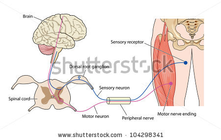
Peripheral nerves are the nerves that extend beyond the spinal cord to every other part of the body. They send impulses from the brain and spinal cord to our various organs and tissues and relay information back to the central nervous system in a complex interplay that enables us to function. Damage to peripheral nerves interferes with this communication process and can cause a wide range of problems.
Peripheral nerves are vulnerable to injury by many means. Diabetes and traumatic or repetitive strain injury are among the most common causes of peripheral nerve damage. Infections, autoimmune disorders, exposure to toxins, nutritional deficiencies, and hereditary conditions are among myriad other causes of neuropathy.
Peripheral neuropathies can affect motor neurons (which control intentional muscle movement), sensory neurons (which provide feedback to the brain from the muscles), and autonomic neurons (which control the muscles required for breathing, swallowing and other autonomic functions). Motor-nerve damage can lead to many mobility-limiting problems with the muscles, such as painful cramping, involuntary twitching, weakness, wasting, and complete or partial paralysis. Damage to sensory neurons can also affect mobility, due to sensations of numbness or pain and the loss of balance and sense of position. Such damage makes it difficult to coordinate the movements required to walk, get dressed, and perform other activities of daily living.
Some forms of peripheral nerve damaged can be managed with medication and/or physical therapy; others cannot. Approaches to treatment and research vary widely depending on the cause and type of damage. Mobility Project researchers are working on a novel strategy to restore hand movement in people with certain peripheral nerve injuries.
Clinical manifestations
The various clinical presentations of patients with peripheral nerve injuries are obviously dependent on the particular nerve or nerves that have been injured as well as the severity and level of the lesion. It is the case with the majority of traumatic peripheral nerve injuries that pain and loss of sensory and motor function are noted either immediately or within days of the inciting traumatic event.
Brachial plexus injuries. The clinical manifestations of brachial plexus injuries are particularly dependent on the specific elements of the plexus that have been injured. The plexus originates from the roots of C5 through T1. For the most part, the C5 root generally gives rise to deltoid function, the C6 root to biceps function, the C7 root to triceps function, the C8 root to the deep flexors of the hand and forearm, and the T1 root to the intrinsic muscle function of the hand. Using these motor functions as a guideline, along with the appropriate sensory distributions, the extent of injury can often be determined by clinical examination.
Median nerve injuries. The most significant and debilitating clinical manifestation of a median nerve injury is loss of pinch and grip strength secondary to denervation of the flexor pollicis longus muscle and the intrinsic muscles of the thenar eminence. A median nerve injury occurring high in its course through the arm will also decrease pronation strength, result in atrophy of forearm muscle mass, and cause sensation deficit of both the volar and palmar aspects of the lateral 3.5 digits.
Radial nerve injuries. The radial nerve originates in the distal axilla and is susceptible at that site to compression injuries, which produce “Saturday night palsy.” The hallmark of such a proximal radial nerve injury is the patient’s inability to extend the forearm secondary to loss of triceps innervation. The majority of radial nerve injuries occur at the midhumeral level secondary to fracture and present with sparing of triceps muscle function in the face of more distal radial distribution loss such as a wrist drop and inability to extend the fingers and thumb. Typically, the sensory deficits involved with radial nerve injury are minimal, involving only the dorsum of the hand and the anatomical snuffbox region.
Ulnar nerve injuries. The ulnar nerve receives its entire contribution from the medial cord of the brachial plexus. In an injury producing a total ulnar palsy, a patient will present with a classic hand posture in which there is partial flexion or “clawing” of the little finger and, to a lesser extent, the ring finger. In the case of a compression syndrome, sensory symptoms in the form of paresthesias and numbness in the little and ring fingers may be the patient’s first complaint. With time, patients will experience atrophy of the intrinsic muscles of the hand, producing a characteristic appearance and significant hand weakness.
Spinal accessory nerve injuries. In 1993, Donner and Kline found the 2 most common complaints of 83 patients with an extracranial spinal accessory nerve injury to be the inability to raise the arm above the horizontal plane and a “dragging pain” in the shoulder. The inability to raise the arm was secondary to a degree of trapezius weakness (particularly the upper trapezius) seen in all patients, whereas the dragging pain was thought to be due to downward tension on the shoulder joint secondary to chronic sagging of the shoulder. Also, on physical exam, scapular winging is a frequent finding distinguishable from the winging of serratus anterior palsy by its disappearance when the patient’s arm is raised forward, fully extended, and pushed against resistance.
Sciatic nerve injuries. Injury to the sciatic nerve at the proximal buttock level or more distally at the midthigh level can produce various symptoms depending on the amount of functional loss in the peroneal and tibial divisions of the nerve. The peroneal division lies posteriorly within the sciatic nerve making it prone to injury from injections or hip fractures. Patients will present with a foot drop secondary to loss of dorsiflexion and eversion of the foot as well as an inability to extend the toes. Injury to the tibial division of the sciatic produces loss of plantar flexion and inversion of the foot as well as loss of toe flexion. Patients with primarily tibial division injury complain mostly of a burning, hyperesthetic sole of the foot.
Femoral nerve injuries. The femoral nerve is the largest branch of the lumbar plexus, arising from the anterior divisions of L2, L3, and L4 spinal nerves. Injury to this nerve within the pelvis will produce a loss of hip flexion due to denervation of the iliopsoas muscle and a loss of knee extension secondary to denervation of the quadriceps muscles. Patients will find it difficult, if not impossible, to climb stairs with this injury. Injury to the femoral nerve at the level of the pelvis will produce a variable loss of hip flexion and variable sensory loss in the anteromedial thigh; however, absence of quadriceps function with loss of the patellar reflex will be uniform. Thigh-level injury of the femoral nerve within the region of the femoral triangle will also produce a complete loss of quadriceps function.
Peripheral Nerve Injury Rehabilitation
A physical medicine and rehabilitation (PM&R) physician, or physiatrist, often follows patients with peripheral nerve injuries as they go through the phases of healing, whether or not they require surgery. These specialists treat pain, if present, and help to minimize the functional deficits that can develop as a result of a peripheral nerve injury.
Pain is treated by a combination of means, including medication. The most effective medications interfere with pain nerve transmission, and are helpful in decreasing this purposeless output of injured nerves as they heal. Other pain treatments include injections of anesthetics, steroids, or other agents, and new technological devices that emit low-frequency signals masking pain nerve signals. Most types of pain can be greatly minimized by a combination of these means.
Preserving function after a peripheral nerve injury follows a careful examination which details the present capability of an affected area, as well as any vulnerable areas as a result of weakness or disuse. Range of motion of a joint within an affected area can be permanently lost if it is not maintained—even if nerve function is subsequently regained. Lack of joint protection can result in future traumatic arthritis, and thus pain. Modern technology has a furnished a number of assistive devices that can provide temporary independence, while safely protecting joints and zones of nerve healing. In addition, other well functioning areas can be helped to compensate for affected areas.
At Rehab Experts our physical therapists are specialized in the evaluation and treatment of nerve conditions. We develop individual treatment plans that are highly effective and we work with your physician for optimal results.
Reference:
Atlantic Mobility Action Project http://www.amap.ca/index.php?option=com_content&view=article&id=75&Itemid=72
Neurology Medlink http://www.medlink.com/medlinkcontent.asp
Columbia University Medical Center http://www.columbianeurosurgery.org/specialties/peripheral-nerve/treatment/peripheral-nerve-injury-rehabilitation/

